Cancer Diagnostic Imaging

Advanced Imaging to Improve Early Detection & Treatment of Disease

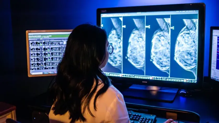
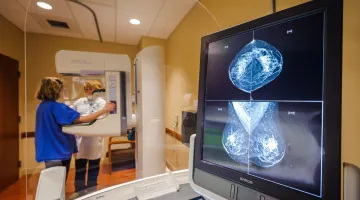
Advances in diagnostic imaging now make it possible to identify and treat diseases in ways that were unimaginable as recently as 20 years ago. South Shore Health's team of board-certified radiologists is the largest in the region and offers patients a full range of diagnostic radiology and imaging services, including a women's diagnostic imaging center on the first floor of the Cancer Center. All diagnostic procedures are fully accredited by the American College of Radiology.

Imaging tests help physicians detect and diagnose disease, make appropriate treatment recommendations, and even monitor your response to therapy. Our goal is to partner with you to obtain the best information possible to make informed diagnostic and treatment recommendations.
 
Because every test is unique and provides different information, imaging test are not always interchangeable. Therefore, while some tests—like x-rays and computed tomography (CT) scans—use radiation to capture images of the body, others—like ultrasound and magnetic resonance imaging (MRI), do not. Tests are never ordered unless the benefit of having the test, outweighs any potential risk. If a recommended test requires use of radiation, our doctors will make every effort to reduce radiation dose as much as possible without compromising the image quality needed for evaluation.

To make an appointment with diagnostic imaging, dial (781) 624-4090. 
 

Mammography uses a low-dose x-ray system to create images of the breast that so doctors can detect cancer. Recent advances in technology have helped enhance the images produced. For example, digital mammography captures images of the breast that can be seen on a computer screen, and computer-aided detection (CAD) software can search digitized mammographic images for abnormal areas of the breast that require further analysis. Learn more. 
Breast Tomosynthesis (TOMO) is a mammography system that creates a series of three-dimensional images of the breast that improves the physician’s ability to detect breast cancer and helps reduce callbacks for additional imaging. 
 

A bone scan is used to examine the bones for damage caused by cancer that either started there or that has spread from another part of the body. A small amount of radioactive material is injected into a vein and after it collects in the bones, a scanner will detect it and create detailed images of the bones on a computer screen.

Brigham and Women's Harbor Medical Associates provides computed tomography (CT) scans. Using a special machine, CT scan uses x-rays to create a series of detailed pictures of areas inside the body taken from different angles. The three-dimensional images reveal abnormalities in both bone and soft tissues, including organs, muscles, and tumors. For some CT exams, a special material called a contrast dye is used to make the area of the body being studied easier to see.

A special (low dose radiation) CT scan may be used as a screening tool for some individuals at high risk of developing lung cancer.

This safe and painless procedure produces pictures of the inside of the body using high-frequency sound waves. The sound waves are transmitted through a probe that is placed directly on the skin over the area that needs to be imaged. Because the images, called sonograms, are captured in real time, they can show the structure and movement of the body’s internal organs, as well as blood flowing through blood vessels.

Nuclear medicine tests use small amounts of radioactive material to diagnose disease. The radioactive substance is injected into the body, locates specific cells or tissues―including cancer cells―and binds to them. Special machines (such as a PET scanner) create images that detect the radioactive substance and help physicians find cancer, see how far it has spread, or assess how well a treatment is working.

This common, noninvasive test involves exposing an area to a small dose of radiation to create pictures of the inside of the body.

Diagnostic Imaging News

Dr. Jabbar
Blog

Bone Health for Survivorship

Bone Health for Survivorship

A view of a 3D mammography scanner
Blog

Three Facts about 3D Mammography

Three Facts about 3D Mammography

A view of a 3D mammography scanner
Blog

The Importance of Maintaining Regular Health Screenings

The Importance of Maintaining Regular Health Screenings